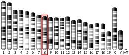
Rex1
Rex1 (Zfp-42) is a known marker of pluripotency, and is usually found in undifferentiated embryonic stem cells. In addition to being a marker for pluripotency, its regulation is also critical in maintaining a pluripotent state.[5] As the cells begin to differentiate, Rex1 is severely and abruptly downregulated.[6]
Discovery
Rex1 was discovered by Hosler, BA et al. in 1989 when studying F9 murine teratocarcinoma stem cells. They found that these teratocarcinoma stem cells expressed high levels of Rex1, and that they resembled pluripotent stem cells of the inner cell mass (ICM).[7] Hosler, BA et al. found that these teratocarcinoma stem cells, when in the presence of retinoic acid (RA), differentiated into nontumorigenic cells resembling extraembryonic endoderm of early mouse embryos.[8] They were able to isolate the nucleotide sequence for Rex1 using differential hybridization of an F9 cell. They named it Rex1 for reduced expression 1 because there was a steady decline of its mRNA levels within 12 hours of the addition of RA.[8]
Structure
Rex1 is a protein that in humans is encoded by the ZFP42 gene.[9][10] The Rex1 protein is 310 amino acids long, and has four closely spaced zinc fingers at 188–212, 217–239, 245–269, and 275–299.[7]
p38 MAPK & Mesenchymal Stem Cells
Rex1 has been found to be critically important in maintaining proliferative state in mesenchymal stem cells (MSC), while simultaneously preventing differentiation. Both umbilical cord blood MSC and adipose MSC express high levels of Rex1, while bone marrow MSC expressed low levels of Rex1. Proliferation rates are highly correlated with Rex1 expression levels, meaning high Rex1 expression is correlated with high levels of proliferation. The MSCs with weak Rex1 expression, have activated p38 MAPK and high expression levels of MKK3. Thus, Rex1 expression is inversely correlated with p38 MAPK activation, and positively correlated with high proliferation rates.[11] Rex1 was found to inhibit MKK3 expression, which activates p38 MAPK. Activated p38 MAPK, in turn, inhibits proliferation. Rex1 was also found to inhibit NOTCH and STAT3, two transcription factors which lead to differentiation.[11] Therefore, Rex1 expression allows for high levels of proliferation, and prevents differentiation through a network of various transcription factors and protein kinases.
Embryo Development
Tissue Derivation
During embryogenesis, the inner cell mass (ICM) is separated from the trophoblast. The stem cells derived from the ICM and trophectoderm have been found to express high levels of Oct3/4 and Rex1.[12] As the ICM matures and begins to form the epiblast, and primitive ectoderm, the cells in the ICM have been found to be a heterogenous population, with varying levels of Rex1 expression. Rex1−/Oct3/4− triggers trophectoderm differentiation, while Rex1+/Oct3/4+ cells predominantly differentiate into primitive endoderm and mesoderm.[13] Also, Rex1−/Oct3/4+ cells differentiate into cells of primitive ectoderm, the somatic cell lineage.[14]
Gene Control
Studies have shown that PEG3 and Nespas are downstream targets of Rex1.[15] Rex1 can control the expression of Peg3 via epigenetic changes. YY1 has been shown to be involved in setting up DNA methylation on the maternal allele of PEG3 during oogenesis.[16] Rex1 was found to protect the paternal allele from being methylated, and keep the PEG3 gene unmethylated during early embryogenesis.[15] Rex1 exhibits gene control in developing embryos via its epigenetic control on genes such as PEG3, which has been identified as playing a key role in fetal growth rates [17]
Expression in Adult Tissues
The only adult tissue Rex1 has been identified in are the testicles. Using in situ hybridization it was determined that the spermatocytes in the more inner layers of the testicles are expressing Rex1.[18] Thus, the male germ cells undergoing meiosis are the specific cells in the testicles that express Rex1. It has not been observed, however, that Rex1 is expressed in the female germ cells.
Rex1 Interactions with Other Transcription Factors
Rex1 participates in a network of transcription factors that all work to regulate each other via varying expression levels.
Nanog
The Nanog protein has been found to be a transcriptional activator for the Rex-1 promoter, playing a key role in sustaining Rex1 expression. Knockdown of Nanog in embryonic stem cells results in a reduction of Rex-1 expression, while forced expression of Nanog stimulates Rex-1 expression.[5] Nanog regulates the transcription of Rex1 through 2 strong transactivation domains on the C-terminus which are required to activate the Rex1 promoter.[5]
NOTCH
Rex1 has been found to inhibit the expression of NOTCH, thus preventing differentiation.[11]
STAT3
Rex1 has been found to inhibit the expression of STAT3, thus preventing differentiation.[11]
Sox2
Cooperative regulation of Rex1 is seen with Sox2 and Nanog.[5]
Oct3/4
Oct3/4 can both repress and activate the Rex1 promoter. In cells that already express high level of Oct3/4, exogenously transfected Oct3/4 will lead to the repression of Rex1.[19] However, in cells that are not actively expressing Oct3/4, an exogenous transfection of Oct3/4 will lead to the activation of Rex1.[19] This implies a dual regulatory ability of Oct3/4 on Rex1. At low levels of the Oct3/4 protein, the Rex1 promoter is activated, while at high levels of the Oct3/4 protein, the Rex1 promoter is repressed.
References
- ^ a b c GRCh38: Ensembl release 89: ENSG00000179059 – Ensembl, May 2017
- ^ a b c GRCm38: Ensembl release 89: ENSMUSG00000051176 – Ensembl, May 2017
- ^ "Human PubMed Reference:". National Center for Biotechnology Information, U.S. National Library of Medicine.
- ^ "Mouse PubMed Reference:". National Center for Biotechnology Information, U.S. National Library of Medicine.
- ^ a b c d Shi W, Wang H, Pan G, Geng Y, Guo Y, Pei D (August 2006). "Regulation of the pluripotency marker Rex-1 by Nanog and Sox2". The Journal of Biological Chemistry. 281 (33): 23319–23325. doi:10.1074/jbc.M601811200. PMID 16714766.
- ^ Wang J, Rao S, Chu J, Shen X, Levasseur DN, Theunissen TW, Orkin SH (November 2006). "A protein interaction network for pluripotency of embryonic stem cells". Nature. 444 (7117): 364–368. Bibcode:2006Natur.444..364W. doi:10.1038/nature05284. PMID 17093407. S2CID 4404796.
- ^ a b "ZooMAb® Antibodies Frequently Asked Questions".
- ^ a b Hosler BA, LaRosa GJ, Grippo JF, Gudas LJ (December 1989). "Expression of REX-1, a gene containing zinc finger motifs, is rapidly reduced by retinoic acid in F9 teratocarcinoma cells". Molecular and Cellular Biology. 9 (12): 5623–5629. doi:10.1128/mcb.9.12.5623. PMC 363733. PMID 2511439.
- ^ Henderson JK, Draper JS, Baillie HS, Fishel S, Thomson JA, Moore H, Andrews PW (Jul 2002). "Preimplantation human embryos and embryonic stem cells show comparable expression of stage-specific embryonic antigens". Stem Cells. 20 (4): 329–337. doi:10.1634/stemcells.20-4-329. PMID 12110702. S2CID 9239873.
- ^ "Entrez Gene: ZFP42 zinc finger protein 42 homolog (mouse)".
- ^ a b c d Bhandari DR, Seo KW, Roh KH, Jung JW, Kang SK, Kang KS (May 2010). Pera M (ed.). "REX-1 expression and p38 MAPK activation status can determine proliferation/differentiation fates in human mesenchymal stem cells". PLOS ONE. 5 (5): e10493. Bibcode:2010PLoSO...510493B. doi:10.1371/journal.pone.0010493. PMC 2864743. PMID 20463961.
- ^ Garcia-Tuñon I, Guallar D, Alonso-Martin S, Benito AA, Benítez-Lázaro A, Pérez-Palacios R, et al. (July 2011). "Association of Rex-1 to target genes supports its interaction with Polycomb function". Stem Cell Research. 7 (1): 1–16. doi:10.1016/j.scr.2011.02.005. PMID 21530438.
- ^ Niwa H, Miyazaki J, Smith AG (April 2000). "Quantitative expression of Oct-3/4 defines differentiation, dedifferentiation or self-renewal of ES cells". Nature Genetics. 24 (4): 372–376. doi:10.1038/74199. PMID 10742100. S2CID 33012290.
- ^ Toyooka Y, Shimosato D, Murakami K, Takahashi K, Niwa H (March 2008). "Identification and characterization of subpopulations in undifferentiated ES cell culture". Development. 135 (5): 909–918. doi:10.1242/dev.017400. PMID 18263842. S2CID 24530633.
- ^ a b Kim JD, Kim H, Ekram MB, Yu S, Faulk C, Kim J (April 2011). "Rex1/Zfp42 as an epigenetic regulator for genomic imprinting". Human Molecular Genetics. 20 (7): 1353–1362. doi:10.1093/hmg/ddr017. PMC 3049358. PMID 21233130.
- ^ Kim JD, Kang K, Kim J (September 2009). "YY1's role in DNA methylation of Peg3 and Xist". Nucleic Acids Research. 37 (17): 5656–5664. doi:10.1093/nar/gkp613. PMC 2761279. PMID 19628663.
- ^ Curley JP, Barton S, Surani A, Keverne EB (June 2004). "Coadaptation in mother and infant regulated by a paternally expressed imprinted gene". Proceedings. Biological Sciences. 271 (1545): 1303–1309. doi:10.1098/rspb.2004.2725. PMC 1691726. PMID 15306355.
- ^ Rogers MB, Hosler BA, Gudas LJ (November 1991). "Specific expression of a retinoic acid-regulated, zinc-finger gene, Rex-1, in preimplantation embryos, trophoblast and spermatocytes". Development. 113 (3): 815–824. doi:10.1242/dev.113.3.815. PMID 1821852.
- ^ a b Ben-Shushan E, Thompson JR, Gudas LJ, Bergman Y (April 1998). "Rex-1, a gene encoding a transcription factor expressed in the early embryo, is regulated via Oct-3/4 and Oct-6 binding to an octamer site and a novel protein, Rox-1, binding to an adjacent site". Molecular and Cellular Biology. 18 (4): 1866–1878. doi:10.1128/mcb.18.4.1866. PMC 121416. PMID 9528758.
Further reading
- Brandenberger R, Wei H, Zhang S, Lei S, Murage J, Fisk GJ, et al. (June 2004). "Transcriptome characterization elucidates signaling networks that control human ES cell growth and differentiation". Nature Biotechnology. 22 (6): 707–716. doi:10.1038/nbt971. PMID 15146197. S2CID 27764390.
- Moriscot C, de Fraipont F, Richard MJ, Marchand M, Savatier P, Bosco D, et al. (April 2005). "Human bone marrow mesenchymal stem cells can express insulin and key transcription factors of the endocrine pancreas developmental pathway upon genetic and/or microenvironmental manipulation in vitro". Stem Cells. 23 (4): 594–603. doi:10.1634/stemcells.2004-0123. PMID 15790780. S2CID 3262995.
- Mongan NP, Martin KM, Gudas LJ (December 2006). "The putative human stem cell marker, Rex-1 (Zfp42): structural classification and expression in normal human epithelial and carcinoma cell cultures". Molecular Carcinogenesis. 45 (12): 887–900. doi:10.1002/mc.20186. PMID 16865673. S2CID 23613555.
- Roche S, Richard MJ, Favrot MC (May 2007). "Oct-4, Rex-1, and Gata-4 expression in human MSC increase the differentiation efficiency but not hTERT expression" (PDF). Journal of Cellular Biochemistry. 101 (2): 271–280. doi:10.1002/jcb.21185. PMID 17211834. S2CID 44892102.
- Rezende NC, Lee MY, Monette S, Mark W, Lu A, Gudas LJ (August 2011). "Rex1 (Zfp42) null mice show impaired testicular function, abnormal testis morphology, and aberrant gene expression". Developmental Biology. 356 (2): 370–382. doi:10.1016/j.ydbio.2011.05.664. PMC 3214085. PMID 21641340.


