Vitelline duct
| Vitelline duct | |
|---|---|
 | |
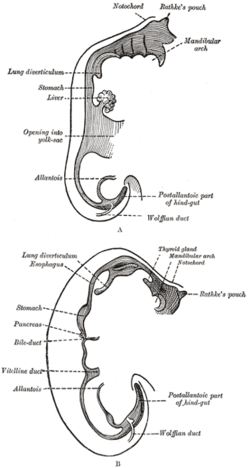
 Sketches in profile of two stages in the development of the human digestive tube. (Vitelline duct labeled on bottom image.) | |
| Details | |
| Days | 28 |
| Precursor | Midgut, yolk sac |
| Identifiers | |
| Latin | ductus vitellinus |
| MeSH | D014816 |
| Anatomical terminology | |
In the human embryo, the vitelline duct, also known as the vitellointestinal duct,[1] the yolk stalk,[1] the omphaloenteric duct,[1] or the omphalomesenteric duct,[1] is a long narrow tube that joins the yolk sac to the midgut lumen of the developing fetus.[2] It appears at the end of the fourth week, when the yolk sac (also known as the umbilical vesicle) presents the appearance of a small pear-shaped vesicle.
Function
Obliteration
Generally, the duct fully obliterates (narrows and disappears) during the 5–6th week of fertilization age (9th week of gestational age), but a failure of the duct to close is termed a vitelline fistula. This results in discharge of meconium from the navel (umbilicus).[2] About two percent of fetuses exhibit a type of vitelline fistula characterized by persistence of the proximal part of the vitelline duct as a diverticulum protruding from the small intestine, Meckel's diverticulum, which is typically situated within two feet of the ileocecal junction and may be attached by a fibrous cord to the abdominal wall at the umbilicus.
Persistence
The yolk sac can be seen in the afterbirth as a small, somewhat oval-shaped body, the diameter of which varies from 1 mm to 5 mm. It is situated between the amnion and the chorion and may lie on the placenta or at a varying distance from it.
Clinical significance
Meckel's diverticulum
Sometimes a narrowing of the lumen of the ileum is seen opposite the site of attachment of the duct. On this site of attachment, sometimes a pathological Meckel's diverticulum may be present.
A mnemonic used to recall details of a Meckel's diverticulum is as follows: "2 inches long, within 2 feet of ileocecal valve, 2 times as common in males than females, 2% of population, 2% symptomatic, 2 types of ectopic tissue: gastric and pancreatic". In the decades since the mnemonic was developed, further epidemiology has found the incidence of symptomatic diverticulae to be 4%, not 2%,[3][4] and the incidence to be 2–5x greater in males than females, but the mnemonic is still helpful.
Additional images
-
Front view of two successive stages in the development of the digestive tube.
References
- ^ a b c d Elsevier, Dorland's Illustrated Medical Dictionary, Elsevier.
- ^ a b Le, Tao; Bhushan, Vikas; Vasan, Neil (2010). First Aid for the USMLE Step 1: 2010 20th Anniversary Edition. USA: The McGraw-Hill Companies, Inc. pp. 122. ISBN 978-0-07-163340-6.
- ^ Robbins and Cotran, Pathologic Basis of Disease, 8th ed., p. 766
- ^ Brant and Helms, Fundamentals of Diagnostic Radiology, 4th ed., p. 778
Further reading
- WebMD (2009). "omphalomesenteric duct". Webster's New World Medical Dictionary (3rd ed.). Houghton Mifflin Harcourt. pp. 305–6. ISBN 978-0-544-18897-6.
