Src (遺伝子)
がん原遺伝子チロシンプロテインキナーゼSrc(Proto-oncogene tyrosine-protein kinase Src)は、ヒトにおいてSRC遺伝子にコードされる非受容体型チロシンキナーゼタンパク質である。がん原遺伝子c-Srcあるいは単にc-Srcとしても知られている。このタンパク質は他のタンパク質の特定のチロシン残基をリン酸化する。c-Srcチロシンキナーゼの活性の上昇は、他のシグナルを促進することによってがんの進行と関連していることが示唆されている[5]。c-SrcはSH2ドメイン、SH3ドメイン、チロシンキナーゼドメインを含んでいる。
c-Srcは、細胞性Srcキナーゼ(cellular Src kinase)の略であり、C末端Srcキナーゼ(C-terminal Src kinase、CSK)と混同してはならない。CSKはc-SrcのC末端をリン酸化し、Srcを不活性にする酵素である。c-Srcは非受容体型チロシンキナーゼ (nRTKs)の中で広く研究されている酵素である。
Src(サルコーマ〔sarcoma; 肉腫〕の短縮形であるため、サークと発音される)は、J・マイケル・ビショップとハロルド・ヴァーマスによって発見されたチロシンキナーゼをコードするがん原遺伝子である。この業績によってビショップとヴァーマスは1989年のノーベル生理学・医学賞を受賞した[6]。c-SrcはSrcファミリーキナーゼと呼ばれる非受容体型チロシンキナーゼのファミリーに属する。
この遺伝子は、ラウス肉腫ウイルスのv-Src遺伝子に似ている。このがん遺伝子は胚発生および細胞成長を制御する役割を果たしている。この遺伝子にコードされているタンパク質はチロシンキナーゼであり、その活性はCskによるリン酸化によって阻害される。この遺伝子の変異は、結腸癌の悪性化に関与している。この遺伝子関して同じタンパク質をコードする2種類の転写変異体が見付かっている[7]。
発見
1979年、J・マイケル・ビショップとハロルド・ヴァーマスは、正常なニワトリがv-Srcと構造的に近縁関係にある遺伝子を含むことを発見した[8]。この正常な細胞遺伝子はc-src(細胞性src; cellular-src)と呼ばれた[9]。この発見は、がんが外的な物質(ウイルス遺伝子)によって引き起こされるというモデルから、細胞中に正常に存在する遺伝子ががんを引き起こすというモデルへと、がんに関する考え方を変化させた。現在は、ある時点において、祖先ウイルスがその細胞ホストのc-Src遺伝子を誤って組み込んだと考えられている。そのうち、この正常遺伝子は、ラウス肉腫ウイルス内で異常に機能するがん遺伝子へと変異した。がん遺伝子をニワトリに導入すると、がんが引き起こされる。
構造および機能
Srcファミリーキナーゼには、c-Src、YES1、FYN、FGR、LYN、BLK、HCK、Lckの9種類が存在する[10]。これらのSrcファミリーの発現は、全ての組織ならびに細胞種全体で同じではない。Src、Fyn、Yesは、全ての細胞種で遍在的に発現しているが、その他は造血細胞において一般に見られる[11][12][13][14]。
c-Srcは、Srcホモロジー (SH) 4ドメイン(SH4ドメイン)、固有領域、SH3ドメイン、SH2ドメイン、触媒ドメイン、短い調節末端の6つの機能領域からなる。Srcが不活性状態の時、527番目のリン酸化チロシン基はSH2ドメインと相互作用し、これがSH3ドメインとリンカードメインの相互作用を助けることによって、しっかり結合した不活性ユニットが保たれる。c-Srcの活性化は、チロシン527の脱リン酸化を引き起こし、これによって構造が不安定化し、SH3ドメイン、SH2ドメイン、キナーゼドメインが広がり、チロシン416がリン酸化される[15][16][17][17]。
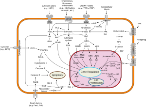
c-Srcは接着受容体、受容体型チロシンキナーゼ、Gタンパク質共役受容体、サイトカイン受容体を含む多くの膜貫通タンパク質によって活性化される。ほとんどの研究は受容体型チロシンキナーゼについて調べており、これらの例としては血小板由来増殖因子受容体 (PDGFR) 経路や上皮成長因子受容体 (EGFR) がある。srcが活性化されると、生存や血管新生、増殖、浸潤経路を促進する。
がんにおける役割
c-Src経路の活性化は、結腸、肝臓、肺、乳房、膵臓の腫瘍のおよそ50%で観察されている[18]。c-Srcの活性化は生存や血管新生、増殖、浸潤経路を促進するため、がんにおける腫瘍の異常成長が観察される。共通の機構は、c-Srcの持続的活性化を引き起こすc-Srcの活性上昇あるいは過剰発現をもたらす遺伝子変異である。
結腸がん
c-Srcの活性は結腸がんにおいて最もよく特徴付けられている。研究者らは、Srcの発現が前がんポリープにおいて正常粘膜よりも5倍から8倍高いことを明らかにしている[19][20][21]。c-Srcレベルの上昇は、腫瘍の進行ステージや腫瘍の大きさ、腫瘍の悪性度と関連していることも明らかにされている[22][23]。
乳がん
EGFRはc-Srcを活性化するが、EGFもc-Srcの活性を上昇させる。加えて、c-Srcの過剰発現は、EGFRが媒介する過程の応答を高める。したがって、EGFRとc-Srcはどちらも、互いの効果を増強する。c-Srcの発現レベルの上昇は、正常組織と比較してヒト乳がん組織で見られる[24][25][26]。
ヒト上皮成長因子受容体2 (HER2) の過剰発現は、乳がんにおける予後の悪さと関連している[27][28]。ゆえに、c-Srcは乳がんの悪性化において重要な役割を果たしている。
前立腺がん
SrcファミリーキナーゼのSrc、Lyn、Fgrは悪性前立腺細胞において正常前立腺細胞よりも高度に発現している[29]。初代前立腺細胞をLynの阻害剤であるKRX-123で処理すると、細胞はin vitroで増殖、遊走、浸潤能が低くなる[30]。したがって、チロシンキナーゼ阻害剤の使用は前立腺がんの進行を弱める方法となりうる。
薬剤標的として
c-Srcチロシンキナーゼ(と類縁チロシンキナーゼ)を標的とする数多くのチロシンキナーゼ阻害剤が、治療薬としての使用のために開発されている[31]。注目に値する例が、慢性骨髄性白血病 (CML) ならびにフィラデルフィア染色体陽性 (PH+) 急性リンパ性白血病 (ALL) の治療薬として承認されたダサチニブである[32]。ダサチニブは、非ホジキンリンパ腫、悪性乳がんおよび前立腺がんに対する臨床試験も行われている。臨床試験が行われているその他のチロシンキナーゼ阻害薬としては、ボスチニブ[33]、バフェチニブ、AZD-530、XLl-999、KX01、XL228がある[5]。
相互作用
Srcは以下のシグナル経路と相互作用することが明らかにされている。
生存
血管新生
増殖
運動性
ギャラリー
 |
脚注
- ^ a b c GRCh38: Ensembl release 89: ENSG00000197122 - Ensembl, May 2017
- ^ a b c GRCm38: Ensembl release 89: ENSMUSG00000027646 - Ensembl, May 2017
- ^ Human PubMed Reference:
- ^ Mouse PubMed Reference:
- ^ a b Wheeler DL, Iida M, Dunn EF (July 2009). “The role of Src in solid tumors”. Oncologist 14 (7): 667–78. doi:10.1634/theoncologist.2009-0009. PMC 3303596. PMID 19581523.
- ^ “The Nobel Prize in Physiology or Medicine 1989: J. Michael Bishop, Harold E. Varmus”. Nobelprize.org (1989年10月9日). 2014年5月1日閲覧。 “for their discovery of 'the cellular origin of retroviral oncogenes'”
- ^ “Entrez Gene: SRC v-src sarcoma (Schmidt-Ruppin A-2) viral oncogene homolog (avian)”. 2014年5月1日閲覧。
- ^ Stehelin D, Fujita DJ, Padgett T, Varmus HE, Bishop JM. (1977). “Detection and enumeration of transformation-defective strains of avian sarcoma virus with molecular hybridization”. Virology 76 (2): 675–84. doi:10.1016/0042-6822(77)90250-1. PMID 190771.
- ^ Oppermann H, Levinson AD, Varmus HE, Levintow L, Bishop JM (April 1979). “Uninfected vertebrate cells contain a protein that is closely related to the product of the avian sarcoma virus transforming gene (src)”. Proc. Natl. Acad. Sci. U.S.A. 76 (4): 1804–8. Bibcode: 1979PNAS...76.1804O. doi:10.1073/pnas.76.4.1804. PMC 383480. PMID 221907.
- ^ Thomas SM, Brugge JS (1997). “Cellular functions regulated by Src family kinases”. Annu. Rev. Cell Dev. Biol. 13: 513–609. doi:10.1146/annurev.cellbio.13.1.513. PMID 9442882.
- ^ Cance WG, Craven RJ, Bergman M, Xu L, Alitalo K, Liu ET (December 1994). “Rak, a novel nuclear tyrosine kinase expressed in epithelial cells”. Cell Growth Differ. 5 (12): 1347–55. PMID 7696183.
- ^ Lee J, Wang Z, Luoh SM, Wood WI, Scadden DT (January 1994). “Cloning of FRK, a novel human intracellular SRC-like tyrosine kinase-encoding gene”. Gene 138 (1–2): 247–51. doi:10.1016/0378-1119(94)90817-6. PMID 7510261.
- ^ Oberg-Welsh C, Welsh M (January 1995). “Cloning of BSK, a murine FRK homologue with a specific pattern of tissue distribution”. Gene 152 (2): 239–42. doi:10.1016/0378-1119(94)00718-8. PMID 7835707.
- ^ Thuveson M, Albrecht D, Zürcher G, Andres AC, Ziemiecki A (April 1995). “iyk, a novel intracellular protein tyrosine kinase differentially expressed in the mouse mammary gland and intestine”. Biochem. Biophys. Res. Commun. 209 (2): 582–9. doi:10.1006/bbrc.1995.1540. PMID 7733928.
- ^ Cooper JA, Gould KL, Cartwright CA, Hunter T (March 1986). “Tyr527 is phosphorylated in pp60c-src: implications for regulation”. Science 231 (4744): 1431–4. Bibcode: 1986Sci...231.1431C. doi:10.1126/science.2420005. PMID 2420005.
- ^ Okada M, Nakagawa H (December 1989). “A protein tyrosine kinase involved in regulation of pp60c-src function”. J. Biol. Chem. 264 (35): 20886–93. PMID 2480346.
- ^ a b Nada S, Okada M, MacAuley A, Cooper JA, Nakagawa H (May 1991). “Cloning of a complementary DNA for a protein-tyrosine kinase that specifically phosphorylates a negative regulatory site of p60c-src”. Nature 351 (6321): 69–72. Bibcode: 1991Natur.351...69N. doi:10.1038/351069a0. PMID 1709258.
- ^ Dehm SM, Bonham K (April 2004). “SRC gene expression in human cancer: the role of transcriptional activation”. Biochem. Cell Biol. 82 (2): 263–74. doi:10.1139/o03-077. PMID 15060621.
- ^ Bolen JB, Rosen N, Israel MA (November 1985). “Increased pp60c-src tyrosyl kinase activity in human neuroblastomas is associated with amino-terminal tyrosine phosphorylation of the src gene product”. Proc. Natl. Acad. Sci. U.S.A. 82 (21): 7275–9. Bibcode: 1985PNAS...82.7275B. doi:10.1073/pnas.82.21.7275. PMC 390832. PMID 2414774.
- ^ Cartwright CA, Kamps MP, Meisler AI, Pipas JM, Eckhart W (June 1989). “pp60c-src activation in human colon carcinoma”. J. Clin. Invest. 83 (6): 2025–33. doi:10.1172/JCI114113. PMC 303927. PMID 2498394.
- ^ Talamonti MS, Roh MS, Curley SA, Gallick GE (January 1993). “Increase in activity and level of pp60c-src in progressive stages of human colorectal cancer”. J. Clin. Invest. 91 (1): 53–60. doi:10.1172/JCI116200. PMC 329994. PMID 7678609.
- ^ Aligayer H, Boyd DD, Heiss MM, Abdalla EK, Curley SA, Gallick GE (January 2002). “Activation of Src kinase in primary colorectal carcinoma: an indicator of poor clinical prognosis”. Cancer 94 (2): 344–51. doi:10.1002/cncr.10221. PMID 11900220.
- ^ Cartwright CA, Meisler AI, Eckhart W (January 1990). “Activation of the pp60c-src protein kinase is an early event in colonic carcinogenesis”. Proc. Natl. Acad. Sci. U.S.A. 87 (2): 558–62. Bibcode: 1990PNAS...87..558C. doi:10.1073/pnas.87.2.558. PMC 53304. PMID 2105487.
- ^ Ottenhoff-Kalff AE, Rijksen G, van Beurden EA, Hennipman A, Michels AA, Staal GE (September 1992). “Characterization of protein tyrosine kinases from human breast cancer: involvement of the c-src oncogene product”. Cancer Res. 52 (17): 4773–8. PMID 1380891.
- ^ Biscardi JS, Belsches AP, Parsons SJ (April 1998). “Characterization of human epidermal growth factor receptor and c-Src interactions in human breast tumor cells”. Mol. Carcinog. 21 (4): 261–72. doi:10.1002/(SICI)1098-2744(199804)21:4<261::AID-MC5>3.0.CO;2-N. PMID 9585256.
- ^ Verbeek BS, Vroom TM, Adriaansen-Slot SS, Ottenhoff-Kalff AE, Geertzema JG, Hennipman A, Rijksen G (December 1996). “c-Src protein expression is increased in human breast cancer. An immunohistochemical and biochemical analysis”. J. Pathol. 180 (4): 383–8. doi:10.1002/(SICI)1096-9896(199612)180:4<383::AID-PATH686>3.0.CO;2-N. PMID 9014858.
- ^ Slamon DJ, Clark GM, Wong SG, Levin WJ, Ullrich A, McGuire WL (January 1987). “Human breast cancer: correlation of relapse and survival with amplification of the HER-2/neu oncogene”. Science 235 (4785): 177–82. Bibcode: 1987Sci...235..177S. doi:10.1126/science.3798106. PMID 3798106.
- ^ Slamon DJ, Godolphin W, Jones LA, Holt JA, Wong SG, Keith DE, Levin WJ, Stuart SG, Udove J, Ullrich A (May 1989). “Studies of the HER-2/neu proto-oncogene in human breast and ovarian cancer”. Science 244 (4905): 707–12. Bibcode: 1989Sci...244..707S. doi:10.1126/science.2470152. PMID 2470152.
- ^ Nam S, Kim D, Cheng JQ, Zhang S, Lee JH, Buettner R, Mirosevich J, Lee FY, Jove R (October 2005). “Action of the Src family kinase inhibitor, dasatinib (BMS-354825), on human prostate cancer cells”. Cancer Res. 65 (20): 9185–9. doi:10.1158/0008-5472.CAN-05-1731. PMID 16230377.
- ^ Chang YM, Bai L, Yang I (2002). “Survey of Src activity and Src-related growth and migration in prostate cancer lines”. Proc Am Assoc Cancer Res 62: 2505a.
- ^ Musumeci F, Schenone S, Brullo C, Botta M (April 2012). “An update on dual Src/Abl inhibitors”. Future Med Chem 4 (6): 799–822. doi:10.4155/fmc.12.29. PMID 22530642.
- ^ Breccia M, Salaroli A, Molica M, Alimena G (2013). “Systematic review of dasatinib in chronic myeloid leukemia”. Onco Targets Ther 6: 257–65. doi:10.2147/OTT.S35360. PMC 3615898. PMID 23569389.
- ^ Amsberg GK, Koschmieder S (2013). “Profile of bosutinib and its clinical potential in the treatment of chronic myeloid leukemia”. Onco Targets Ther 6: 99–106. doi:10.2147/OTT.S19901. PMC 3594007. PMID 23493838.
外部リンク
- src Gene - MeSH・アメリカ国立医学図書館・生命科学用語シソーラス
- src-Family Kinases - MeSH・アメリカ国立医学図書館・生命科学用語シソーラス
- Proteopedia SRC - interactive 3D model of the structure of SRC
- Vega geneview
- Src Info with links in the Cell Migration Gateway


