N-カドヘリン
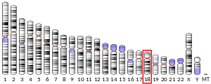
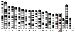
N-カドヘリン(neural cadherin, NCAD)またはカドヘリン2(cadherin-2, CDH2)は、ヒトではCDH2遺伝子にコードされるタンパク質である[5][6]。CD325(cluster of differentiation 325)とも表記される。N-カドヘリンは複数の組織で発現している膜貫通タンパク質であり、細胞間接着を媒介する機能を果たす。心筋においては、N-カドヘリンは介在板に位置するアドヘレンスジャンクションの重要な構成要素である。介在板は、隣接する心筋細胞との機械的・電気的に共役する機能を果たす。N-カドヘリンタンパク質の発現や完全性の変化は、拡張型心筋症などさまざまな疾患で観察される。CDH2遺伝子の変異は症候群性神経発達障害を引き起こすことも特定されている[7]。
構造
N-カドヘリンは99.7 kDa、906アミノ酸からなるタンパク質である[8]。N-カドヘリンはカドヘリンスーパーファミリーに属する古典的(classical)カドヘリンであり、細胞外の5つのカドヘリンリピート、膜貫通領域、高度に保存された細胞質テールから構成される。他のカドヘリンと同様、N-カドヘリンは隣接する細胞上のN-カドヘリンと逆平行型の相互作用を行い、細胞間に線形で接着性の「ジッパー」を形成する[9]。
機能
N-カドヘリンという名称はその神経組織(neural tissue)における役割に由来し、神経細胞、そして後に心筋やがんの転移においても役割を果たしていることが発見された。N-カドヘリンは膜貫通タンパク質で、ホモフィリック(同種分子親和性)結合を行う糖タンパク質であり、カルシウム依存性細胞接着分子ファミリーに属する。こうしたタンパク質には隣接細胞間のホモフィリック相互作用を媒介する細胞外ドメインが存在し、またC末端にはカテニンとの結合を媒介する細胞質テールが存在する。カテニンはアクチン細胞骨格との相互作用を行う[10][11][12]。
発生における役割
N-カドヘリンはカルシウム依存性細胞間接着糖タンパク質として発生に関与しており、原腸形成時に機能する。また左右非対称性の確立にも必要である[13]。
N-カドヘリンは着床後の胚で広く発現しており、中胚葉での高レベルでの発現は成体でも維持される[14]。発生時のN-カドヘリンの変異は原始心臓での細胞接着に最も大きな影響を与え、筋細胞の解離や異常な心臓管の発生が引き起こされる[15]。N-カドヘリンは脊椎動物の心臓発生において、上皮細胞の肉柱への転換や緻密層の形成に関与している[16]。ドミナントネガティブ型N-カドヘリン変異体を発現する筋細胞では筋細胞の分布や心内膜方向への移動に重大な異常が生じ、心筋内での肉柱の形成に欠陥が生じる[17][18]。
心筋における役割
心筋においては、N-カドヘリンは介在板構造に位置する。介在板は隣接する心筋細胞間の末端の連結を行い、機械的・電気的共役を促進する構造である。介在板内にはアドヘレンスジャンクション、デスモソーム、ギャップジャンクションの3種類の接着構造が存在する[19]。N-カドヘリンはアドヘレンスジャンクションの必須の構成要素であり、細胞間接着と筋鞘間の力の伝達を可能にしている[20]。カテニンと複合体を形成したN-カドヘリンは、介在板の機能のマスターレギュレーターである[21]。N-カドヘリンはギャップジャンクションの形成に先立って細胞間結合部に出現し[22][23]、正常な筋線維の形成に重要な役割を果たす[24]。細胞外ドメインに大きな欠失を抱える変異型N-カドヘリンの発現は成体の心室心筋細胞の内在性N-カドヘリンの機能を阻害し、隣接する心筋細胞は細胞間接触やギャップジャンクションプラークを喪失する[25]。
心筋におけるN-カドヘリンの機能の重要性は、遺伝子導入を行ったマウスモデルによって明らかにされている。N-カドヘリンやE-カドヘリンの発現が変化したマウスは拡張型心筋症の表現型を示し、おそらくこれは介在板の機能不全が原因である[26]。心臓特異的タモキシフェン誘導性Cre-N-カドヘリン導入遺伝子によって成体の心臓でN-カドヘリンを除去したマウスでは、介在板の組み立ての異常、拡張型心筋症、心機能不全、サルコメアの長さの減少、Z帯の厚さの増加、コネキシン43の減少、そして筋張力の喪失がみられる。導入遺伝子を発現したマウスは、主に心室頻拍のために2か月以内に死に至る[27]。N-カドヘリンノックアウトマウスを用いたさらなる解析によって、不整脈はイオンチャネルのリモデリングとKv1.5の機能の異常を原因とする可能性が高いことが明らかにされている。これらのマウスモデルでは、活動電位の持続期間の伸長、内向き整流カリウムチャネルの密度の低下、Kv1.5、KCNE2、コルタクチンの発現の低下、筋鞘におけるアクチン細胞骨格の破壊がみられる[28]。
神経における役割
中枢神経系におけるシナプス前後の接着は、少なくともその一部はN-カドヘリンによって媒介されている。N-カドヘリンはカテニンと相互作用して学習と記憶に重要な役割を果たしている。N-カドヘリンの喪失はヒトの注意欠陥・多動性障害(ADHD)、シナプスの機能不全とも関係している[29]。
がんの転移における役割
N-カドヘリンはがん細胞にも広く存在し、経内皮移動の機構となっている。がん細胞が血管の内皮細胞に接着するとSrcキナーゼ経路がアップレギュレーションされ、N-カドヘリンとE-カドヘリンの双方に結合するβ-カテニンのリン酸化が行われる。その結果、2つの隣接する内皮細胞間の連結が解かれ、がん細胞がすり抜けることができるようになる[30]。
臨床的意義
CDH2遺伝子の変異は脳梁、軸索、心臓、目、生殖器の欠陥によって特徴づけられる症候群性神経発達障害を引き起こすことが特定されている[7]。
強迫性障害とトゥレット障害の遺伝的基礎に関する研究では、CDH2の変異はそれ単独では疾患を引き起こす可能性は低いことが示されているが、関連する細胞間接着遺伝子群の一部としてリスク因子となる可能性がある[31]。この点について明確にするためには、より大規模なコホート研究が必要である。
ヒトの拡張型心筋症では、N-カドヘリンの発現が亢進しており、配置が乱雑なものとなっていることが示されている。このことは、心疾患におけるN-カドヘリンタンパク質の組織化の異常が心臓リモデリングの構成因子となっている可能性を示唆している[32]。
相互作用
N-カドヘリン(CDH2)は次に挙げる因子と相互作用することが示されている。
出典
- ^ a b c GRCh38: Ensembl release 89: ENSG00000170558 - Ensembl, May 2017
- ^ a b c GRCm38: Ensembl release 89: ENSMUSG00000024304 - Ensembl, May 2017
- ^ Human PubMed Reference:
- ^ Mouse PubMed Reference:
- ^ “N-cadherin gene maps to human chromosome 18 and is not linked to the E-cadherin gene”. Journal of Neurochemistry 55 (3): 805–12. (September 1990). doi:10.1111/j.1471-4159.1990.tb04563.x. PMID 2384753.
- ^ “Human N-cadherin: nucleotide and deduced amino acid sequence”. Nucleic Acids Research 18 (19): 5896. (October 1990). doi:10.1093/nar/18.19.5896. PMC 332345. PMID 2216790.
- ^ a b “De Novo Pathogenic Variants in N-cadherin Cause a Syndromic Neurodevelopmental Disorder with Corpus Collosum, Axon, Cardiac, Ocular, and Genital Defects”. American Journal of Human Genetics 105 (4): 854–868. (October 2019). doi:10.1016/j.ajhg.2019.09.005. PMC 6817525. PMID 31585109.
- ^ “Protein sequence of human CDH2 (Uniprot ID: P19022)”. Cardiac Organellar Protein Atlas Knowledgebase (COPaKB). 24 September 2015時点のオリジナルよりアーカイブ。20 July 2015閲覧。
- ^ “Structural basis of cell-cell adhesion by cadherins”. Nature 374 (6520): 327–37. (March 1995). Bibcode: 1995Natur.374..327S. doi:10.1038/374327a0. PMID 7885471.
- ^ “Structure and interactions of desmosomal and other cadherins”. Seminars in Cell Biology 3 (3): 157–67. (June 1992). doi:10.1016/s1043-4682(10)80012-1. PMID 1623205.
- ^ “Cadherins: a molecular family important in selective cell-cell adhesion”. Annual Review of Biochemistry 59: 237–52. (1990). doi:10.1146/annurev.bi.59.070190.001321. PMID 2197976.
- ^ “The cytoplasmic domain of the cell adhesion molecule uvomorulin associates with three independent proteins structurally related in different species”. The EMBO Journal 8 (6): 1711–7. (June 1989). doi:10.1002/j.1460-2075.1989.tb03563.x. PMC 401013. PMID 2788574.
- ^ “N-Cadherin, a cell adhesion molecule involved in establishment of embryonic left-right asymmetry”. Science 288 (5468): 1047–51. (May 2000). Bibcode: 2000Sci...288.1047G. doi:10.1126/science.288.5468.1047. PMID 10807574.
- ^ “Dissociated spatial patterning of gap junctions and cell adhesion junctions during postnatal differentiation of ventricular myocardium”. Circulation Research 80 (1): 88–94. (January 1997). doi:10.1161/01.res.80.1.88. PMID 8978327.
- ^ “Developmental defects in mouse embryos lacking N-cadherin”. Developmental Biology 181 (1): 64–78. (January 1997). doi:10.1006/dbio.1996.8443. PMID 9015265.
- ^ “Differential adhesion leads to segregation and exclusion of N-cadherin-deficient cells in chimeric embryos”. Developmental Biology 234 (1): 72–9. (June 2001). doi:10.1006/dbio.2001.0250. PMID 11356020.
- ^ “N-cadherin-catenin interaction: necessary component of cardiac cell compartmentalization during early vertebrate heart development”. Developmental Biology 185 (2): 148–64. (May 1997). doi:10.1006/dbio.1997.8570. PMID 9187080.
- ^ “Trabecular myocytes of the embryonic heart require N-cadherin for migratory unit identity”. Developmental Biology 193 (1): 1–9. (January 1998). doi:10.1006/dbio.1997.8775. PMID 9466883.
- ^ “Spatiotemporal relation between gap junctions and fascia adherens junctions during postnatal development of human ventricular myocardium”. Circulation 90 (2): 713–25. (August 1994). doi:10.1161/01.cir.90.2.713. PMID 8044940.
- ^ “Intercalated discs of mammalian heart: a review of structure and function”. Tissue & Cell 17 (5): 605–48. (1985). doi:10.1016/0040-8166(85)90001-1. PMID 3904080.
- ^ “N-cadherin/catenin complex as a master regulator of intercalated disc function”. Cell Communication & Adhesion 21 (3): 169–79. (June 2014). doi:10.3109/15419061.2014.908853. PMC 6054126. PMID 24766605.
- ^ “Dynamics of early contact formation in cultured adult rat cardiomyocytes studied by N-cadherin fused to green fluorescent protein”. Journal of Molecular and Cellular Cardiology 32 (4): 539–55. (April 2000). doi:10.1006/jmcc.1999.1086. PMID 10756112.
- ^ “[Dynamic assembly of intercalated disc during postnatal development in the rat myocardium]”. Sheng Li Xue Bao 66 (5): 569–74. (October 2014). PMID 25332002.
- ^ “The involvement of adherens junction components in myofibrillogenesis in cultured cardiac myocytes”. Development 114 (1): 173–83. (January 1992). doi:10.1242/dev.114.1.173. PMID 1576958.
- ^ “N-cadherin in adult rat cardiomyocytes in culture. I. Functional role of N-cadherin and impairment of cell-cell contact by a truncated N-cadherin mutant”. Journal of Cell Science 109 ( Pt 1) (1): 1–10. (January 1996). doi:10.1242/jcs.109.1.1. PMID 8834785.
- ^ “Remodeling the intercalated disc leads to cardiomyopathy in mice misexpressing cadherins in the heart”. Journal of Cell Science 115 (Pt 8): 1623–34. (April 2002). doi:10.1242/jcs.115.8.1623. PMID 11950881.
- ^ “Induced deletion of the N-cadherin gene in the heart leads to dissolution of the intercalated disc structure”. Circulation Research 96 (3): 346–54. (February 2005). doi:10.1161/01.RES.0000156274.72390.2c. PMID 15662031.
- ^ “Cortactin is required for N-cadherin regulation of Kv1.5 channel function”. The Journal of Biological Chemistry 286 (23): 20478–89. (June 2011). doi:10.1074/jbc.m111.218560. PMC 3121477. PMID 21507952.
- ^ “CDH2 mutation affecting N-cadherin function causes attention-deficit hyperactivity disorder in humans and mice”. Nature Communications 6187 (12): 625–30. (October 2021). doi:10.1038/s41467-021-26426-1.
- ^ “Multi-scale modelling of cancer cell intravasation: the role of cadherins in metastasis”. Physical Biology 6 (1): 016008. (March 2009). Bibcode: 2009PhBio...6a6008R. doi:10.1088/1478-3975/6/1/016008. PMID 19321920.
- ^ “Rare missense neuronal cadherin gene (CDH2) variants in specific obsessive-compulsive disorder and Tourette disorder phenotypes”. European Journal of Human Genetics 21 (8): 850–4. (August 2013). doi:10.1038/ejhg.2012.245. PMC 3722668. PMID 23321619.
- ^ “Apoptosis-related factors p53, bcl-2 and the defects of force transmission in dilated cardiomyopathy”. Pathology, Research and Practice 206 (9): 625–30. (September 2010). doi:10.1016/j.prp.2010.05.007. PMID 20591580.
- ^ a b c d e “A novel cell-cell junction system: the cortex adhaerens mosaic of lens fiber cells”. Journal of Cell Science 116 (Pt 24): 4985–95. (December 2003). doi:10.1242/jcs.00815. PMID 14625392.
- ^ a b c “N-cadherin-catenin complexes form prior to cleavage of the proregion and transport to the plasma membrane”. The Journal of Biological Chemistry 278 (19): 17269–76. (May 2003). doi:10.1074/jbc.M211452200. PMID 12604612.
- ^ “Identification of plakoglobin domains required for association with N-cadherin and alpha-catenin”. The Journal of Biological Chemistry 270 (34): 20201–6. (August 1995). doi:10.1074/jbc.270.34.20201. PMID 7650039.
- ^ “Densin-180 interacts with delta-catenin/neural plakophilin-related armadillo repeat protein at synapses”. The Journal of Biological Chemistry 277 (7): 5345–50. (February 2002). doi:10.1074/jbc.M110052200. PMID 11729199.
- ^ “Receptor protein tyrosine phosphatase PTPmu associates with cadherins and catenins in vivo”. The Journal of Cell Biology 130 (4): 977–86. (August 1995). doi:10.1083/jcb.130.4.977. PMC 2199947. PMID 7642713.
- ^ “Dynamic interaction of PTPmu with multiple cadherins in vivo”. The Journal of Cell Biology 141 (1): 287–96. (April 1998). doi:10.1083/jcb.141.1.287. PMC 2132733. PMID 9531566.
- ^ “Intracellular substrates of brain-enriched receptor protein tyrosine phosphatase rho (RPTPrho/PTPRT)”. Brain Research 1116 (1): 50–7. (October 2006). doi:10.1016/j.brainres.2006.07.122. PMID 16973135.
- ^ “Lysosomal integral membrane protein 2 is a novel component of the cardiac intercalated disc and vital for load-induced cardiac myocyte hypertrophy”. The Journal of Experimental Medicine 204 (5): 1227–35. (May 2007). doi:10.1084/jem.20070145. PMC 2118572. PMID 17485520.
- ^ “Localization of the novel Xin protein to the adherens junction complex in cardiac and skeletal muscle during development”. Developmental Dynamics 225 (1): 1–13. (September 2002). doi:10.1002/dvdy.10131. PMID 12203715.
関連文献
- “Shared cell adhesion molecule (CAM) homology domains point to CAMs signalling via FGF receptors”. Perspectives on Developmental Neurobiology 4 (2–3): 157–68. (1997). PMID 9168198.
- “Follicular atresia and luteolysis. Evidence of a role for N-cadherin”. Annals of the New York Academy of Sciences 900 (1): 46–55. (2000). Bibcode: 2000NYASA.900...46M. doi:10.1111/j.1749-6632.2000.tb06215.x. PMID 10818391.
- “Cadherin switch in tumor progression”. Annals of the New York Academy of Sciences 1014 (1): 155–63. (April 2004). Bibcode: 2004NYASA1014..155H. doi:10.1196/annals.1294.016. PMID 15153430.
- “N-cadherin as an invasion promoter: a novel target for antitumor therapy?”. Current Opinion in Investigational Drugs 5 (12): 1274–8. (December 2004). PMID 15648948.
- “Extrajunctional distribution of N-cadherin in cultured human endothelial cells”. Journal of Cell Science 102 ( Pt 1) (1): 7–17. (May 1992). doi:10.1242/jcs.102.1.7. PMID 1500442.
- “Plakoglobin, or an 83-kD homologue distinct from beta-catenin, interacts with E-cadherin and N-cadherin”. The Journal of Cell Biology 118 (3): 671–9. (August 1992). doi:10.1083/jcb.118.3.671. PMC 2289540. PMID 1639850.
- “Human N-cadherin: nucleotide and deduced amino acid sequence”. Nucleic Acids Research 18 (19): 5896. (October 1990). doi:10.1093/nar/18.19.5896. PMC 332345. PMID 2216790.
- “N-cadherin gene maps to human chromosome 18 and is not linked to the E-cadherin gene”. Journal of Neurochemistry 55 (3): 805–12. (September 1990). doi:10.1111/j.1471-4159.1990.tb04563.x. PMID 2384753.
- “Expressed cadherin pseudogenes are localized to the critical region of the spinal muscular atrophy gene”. Proceedings of the National Academy of Sciences of the United States of America 92 (9): 3702–6. (April 1995). Bibcode: 1995PNAS...92.3702S. doi:10.1073/pnas.92.9.3702. PMC 42029. PMID 7731968.
- “Structure of the human N-cadherin gene: YAC analysis and fine chromosomal mapping to 18q11.2”. Genomics 22 (1): 172–9. (July 1994). doi:10.1006/geno.1994.1358. PMID 7959764.
- “Expression and localization of N- and E-cadherin in the human testis and epididymis”. International Journal of Andrology 17 (4): 174–80. (August 1994). doi:10.1111/j.1365-2605.1994.tb01239.x. PMID 7995652.
- “Multiple cadherins are expressed in human fibroblasts”. Biochemical and Biophysical Research Communications 235 (2): 355–8. (June 1997). doi:10.1006/bbrc.1997.6707. PMID 9199196.
- “Differential localization of VE- and N-cadherins in human endothelial cells: VE-cadherin competes with N-cadherin for junctional localization”. The Journal of Cell Biology 140 (6): 1475–84. (March 1998). doi:10.1083/jcb.140.6.1475. PMC 2132661. PMID 9508779.
- “Distribution of N-cadherin and NCAM in neurons and endocrine cells of the human embryonic and fetal gastroenteropancreatic system”. Acta Histochemica 100 (1): 83–97. (February 1998). doi:10.1016/s0065-1281(98)80008-1. PMID 9542583.
- “Localization of human cadherin genes to chromosome regions exhibiting cancer-related loss of heterozygosity”. Genomics 49 (3): 467–71. (May 1998). doi:10.1006/geno.1998.5281. PMID 9615235.
- “delta-catenin, an adhesive junction-associated protein which promotes cell scattering”. The Journal of Cell Biology 144 (3): 519–32. (February 1999). doi:10.1083/jcb.144.3.519. PMC 2132907. PMID 9971746.
- “Functional cis-heterodimers of N- and R-cadherins”. The Journal of Cell Biology 148 (3): 579–90. (February 2000). doi:10.1083/jcb.148.3.579. PMC 2174798. PMID 10662782.
- “Proteomic analysis of NMDA receptor-adhesion protein signaling complexes”. Nature Neuroscience 3 (7): 661–9. (July 2000). doi:10.1038/76615. hdl:1842/742. PMID 10862698.
関連項目
- ADH-1
外部リンク
- CDH2 protein, human - MeSH・アメリカ国立医学図書館・生命科学用語シソーラス


